Anatomy of a Foot: Diagrams and Physiology
Your feet carry you through life, but how much do you really know about their anatomy?
Published July 17, 2024

For centuries, Leonardo da Vinci called the human foot "a masterpiece of engineering and a work of art." Yet, in our head-to-heart-centered world, it's often taken for granted.
We cram them into ill-fitting shoes, subject them to punishing workouts like running, and generally ignore them until they scream for attention. Knowing the anatomy of your foot can improve overall musculoskeletal health.
Below, we'll peel the structure, challenge what you think you know about your foundation, and change the way you walk through life—quite literally.
» Start supporting the anatomy of your foot with custom-made orthotics

Meet the Expert
Jasrah Javed holds a master’s degree in musculoskeletal and sports physiotherapy. She is a passionate writer and researcher determined to spread healthcare awareness through her writings and research.
Importance of Understanding Foot Anatomy
When it comes to foot ailments, many of my patients misjudge the location and cause of their pain because they don't understand the anatomy. As a result, they often ignore the problem or end up doctor-shopping.
Being aware of the structure helps you choose appropriate footwear, workout plans, and injury prevention techniques. In my experience as a physiotherapist, many resulting injuries are avoidable. That's why I believe public awareness of foot anatomy can lead to improved quality of life.
» Learn how to overcome foot fractures
Anatomy of the Foot: Bones
Your foot has three parts: the hindfoot, midfoot, and forefoot. All of them are comprised of bones that have different roles:
- Hindfoot: The talus and calcaneus bones are located between the back part of the foot and the ankle joint. They attach the foot to the thigh and cushion it during weight-bearing activities. [1] The resulting flexibility and plasticity help you adjust to uneven terrain.
- Midfoot: The navicular, cuboid, and cuneiform bone are the five tarsal parts that form the middle part of the foot. [2] They arch the foot to help it absorb shock and distribute weight when moving.
- Forefoot: In this section, metatarsal and toe bones (phalanges) work together to maintain stability. [3] These parts propel, support, and flex the foot when walking, running, and leaping. They also help it absorb shock and adjust to different surfaces.

Major Muscles on Top of the Foot
There are many muscles on top of the foot that are vital for stability, mobility, and strength. For example, the abductor hallucis pulls the big toe away from the rest, while the flexor digitorum brevis helps with fine motor control of the toes. [3]
During weight-bearing exercises, these two muscles dynamically modify the shape of the foot to stabilize it. Their synergy also provides proprioceptive input, the body's ability to sense movement, action, and location.
» Check out the best insoles to wear while squatting
On the other hand, some muscles originate in the thigh and pass into the foot, accelerating movement. For instance, the tibialis posterior points it downwards just before the heel strikes during walking. [3] Similarly, the plantar flexor muscles of the gastrocnemius and soleus move the body forward during push-off. [3]
» Discover the best exercises to strengthen the runner's knee

Essential Ligaments and Tendons
Ligaments
Thick bands of tissue called ligaments link the bones, giving them stability and preventing excessive joint movement. One of them is plantar fascia, which is a broad band of tissue that runs down the bottom of the foot. [3] It supports the arch while absorbing shock during walking and running.
The deltoids—on the inner aspect of the ankle—include the tibionavicular, the posterior tibiotalar, the tibiocalcaneal, and the anterior tibiotalar ligament. [3] They connect the tibia and fibula to the talus and calcaneus bones, keeping the foot from sliding outward and preserving the arch.
» Learn more about out-toeing
Next, the calcaneofibular, posterior talofibular, and anterior talofibular ligaments are on the lateral side of the ankle. [1] They also stabilize its joint and prevent excessive inversion—rolling inward—of the foot.
Note: Strong pressures in sports and uneven terrain activities frequently damage these ligaments in the form of ankle sprains.
» Explore the different types of ankle sprains

Tendons
Tendons link muscles and bones, transferring their force during walking or running. One of the most important ones is the Achilles tendon, which helps in rolling the foot downwards (plantarflexion) by joining the calf muscles to the calcaneus. [3]
» Learn the difference between Achilles tendinopathy and tendonitis
Similar to Achilles, the peroneal tendons sustain the foot and ankle during eversion, while the posterior tibial supports the arch and prevents foot inversion, also known as supination.
Another crucial tendon is the posterior tibial, which helps invert the foot and supports the arch. Finally, the peroneal tendons—on the outside of the ankle—also maintain ankle and foot stability while moving. [2]
» Stretch the arch of your foot for pain relief
Critical Functions of the Foot
Shock-Absorption
During weight-bearing activities, the foot and lower leg muscles that are attached to tendons control their position and stabilize the arches. By contracting and relaxing, they also make the upward and downward movement possible.
» Find out what causes pain in the middle part of the foot and how to get rid of it
Your general musculoskeletal health depends on the foot's capacity to support weight and absorb trauma. The three arches—transverse, lateral, and medial longitudinal—are in charge of the process. Plantar fascia and other soft tissues also play a role.
Note: In addition to protecting the foot from injury, shock-absorbing also lowers the chance of impact forces being transmitted up the kinetic chain, reducing the possibility of knee, hip, and spine injury.
Balance and Stability
The arches equally distribute weight over the surface, offering support. Sensors in the foot and ankle also constantly inform the brain about the position and movement, enabling quick corrections to preserve balance.
» Check out the benefits of rolling your feet
Additionally, the lower leg muscles actively support the ankle and joints, especially during activities with sudden direction changes or uneven terrain.

Common Foot Conditions and Pathologies
There are a number of standard foot disorders across all age groups and activity levels:
- Plantar Fasciitis: Defined by inflammation of the thick band of tissue that runs down the bottom of the foot. [4] It's most common in the morning and after extended hours of standing or walking.
- Achilles Tendonitis: Another common problem that causes discomfort and stiffness in the back of the heel due to inflammation of the tendon. [5] This condition is typically made worse by physical activity.
- Bunions: Usually located near the base of the big toe, causing it to deviate towards the second. [6] It results in a bony protrusion on the side of the foot, creating discomfort, swelling, and trouble fitting into shoes.
- Ankle Sprains: Often result from the ligaments supporting the ankle being stretched or torn, sometimes as a result of trauma or a violent twisting action. [7]Possible outcomes are pain, edema, instability, and trouble bearing weight on the injured ankle.
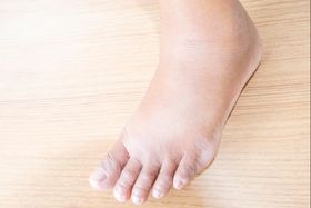
Note: People with collapsed arches or flat feet may also be more susceptible to foot problems. These characteristics change the way the body moves and put more strain on specific parts of it.

How Doctors Treat Common Foot Ailments
Foot pain can have many causes. Healthcare professionals like podiatrists can identify the root of the problem by examining the specific joint or ligament involved.
For example, by looking at the metatarsophalangeal joint—where your big toe connects to your foot—they can diagnose and treat bunions by finding ways to relieve pressure on that area. [8]
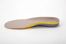
There's a reason you might get this condition. Traditional shoe inserts are a one-size-fits-all solution for a problem that's unique to you. To help you, a doctor would prescribe custom orthotics.
Upstep can help you skip this process with an at-home impression kit that takes minutes. The result? An insole that targets the root cause of your foot pain, providing support and relief. If you don't receive any benefits, you can return them within 180 days for a refund.
Note: Another key to reducing foot pain and discomfort is physical therapy. Exercises that target Achilles tendonitis or ankle sprains relieve pain and restore appropriate foot mechanics. Some examples include stretching, joint mobilization, and massage. [5]

The Key to Healthy Feet and Happy You
Knowing how your foot works (bones, muscles, etc.) helps you appreciate proper alignment and mechanics for activities like walking.
This knowledge empowers you to care for your feet with proper footwear, hygiene, and exercise. Understanding the causes can also help prevent common foot problems and injuries.
By being aware of your foot anatomy, you can take charge of your foot health, leading to better mobility, comfort, and overall well-being. Ready to start?
» Improve your musculoskeletal health with custom-made orthotics
References:
- “Anatomy, bony pelvis and lower limb, foot talus,” PubMed, Jan. 01, 2024. Available: https://pubmed.ncbi.nlm.nih.gov/31082130/
- C. L. Brockett and G. J. Chapman, “Biomechanics of the ankle,” Orthopaedics and Trauma, vol. 30, no. 3, pp. 232–238, Jun. 2016, doi: 10.1016/j.mporth.2016.04.015. Available: https://pubmed.ncbi.nlm.nih.gov/27594929/
- “Anatomy, bony pelvis and lower limb, foot muscles,” PubMed, Jan. 01, 2024. Available: https://pubmed.ncbi.nlm.nih.gov/30969527/
- “Plantar fasciitis,” PubMed, Jan. 01, 2024. Available: https://pubmed.ncbi.nlm.nih.gov/28613727/
- “Achilles tendinopathy,” PubMed, Jan. 01, 2024. Available: https://pubmed.ncbi.nlm.nih.gov/30844176/
- A. S. Aebischer and S. Duff, “Bunions: A review of management,” Australian Journal of General Practice, vol. 49, no. 11, pp. 720–723, Nov. 2020, doi: 10.31128/ajgp-07-20-5541. Available: https://pubmed.ncbi.nlm.nih.gov/33123707/
- G. Vuurberg et al., “Diagnosis, treatment and prevention of ankle sprains: update of an evidence-based clinical guideline,” British Journal of Sports Medicine, vol. 52, no. 15, p. 956, Mar. 2018, doi: 10.1136/bjsports-2017-098106. Available: https://pubmed.ncbi.nlm.nih.gov/29514819/
- K. Han, K. Bae, N. Levine, J. Yang, and J.-S. Lee, “Biomechanical Effect of Foot Orthoses on Rearfoot Motions and Joint Moment Parameters in Patients with Flexible Flatfoot,” Medical Science Monitor, vol. 25, pp. 5920–5928, Aug. 2019, doi: 10.12659/msm.918782. Available: https://pubmed.ncbi.nlm.nih.gov/31393860/
Disclaimer: The information on this blog is for educational purposes only and is not a substitute for professional medical advice.
Upstep does not provide medical diagnosis or treatment. While qualified healthcare professionals create our content, it's essential to consult with your healthcare provider for any foot or ankle concerns you may have.